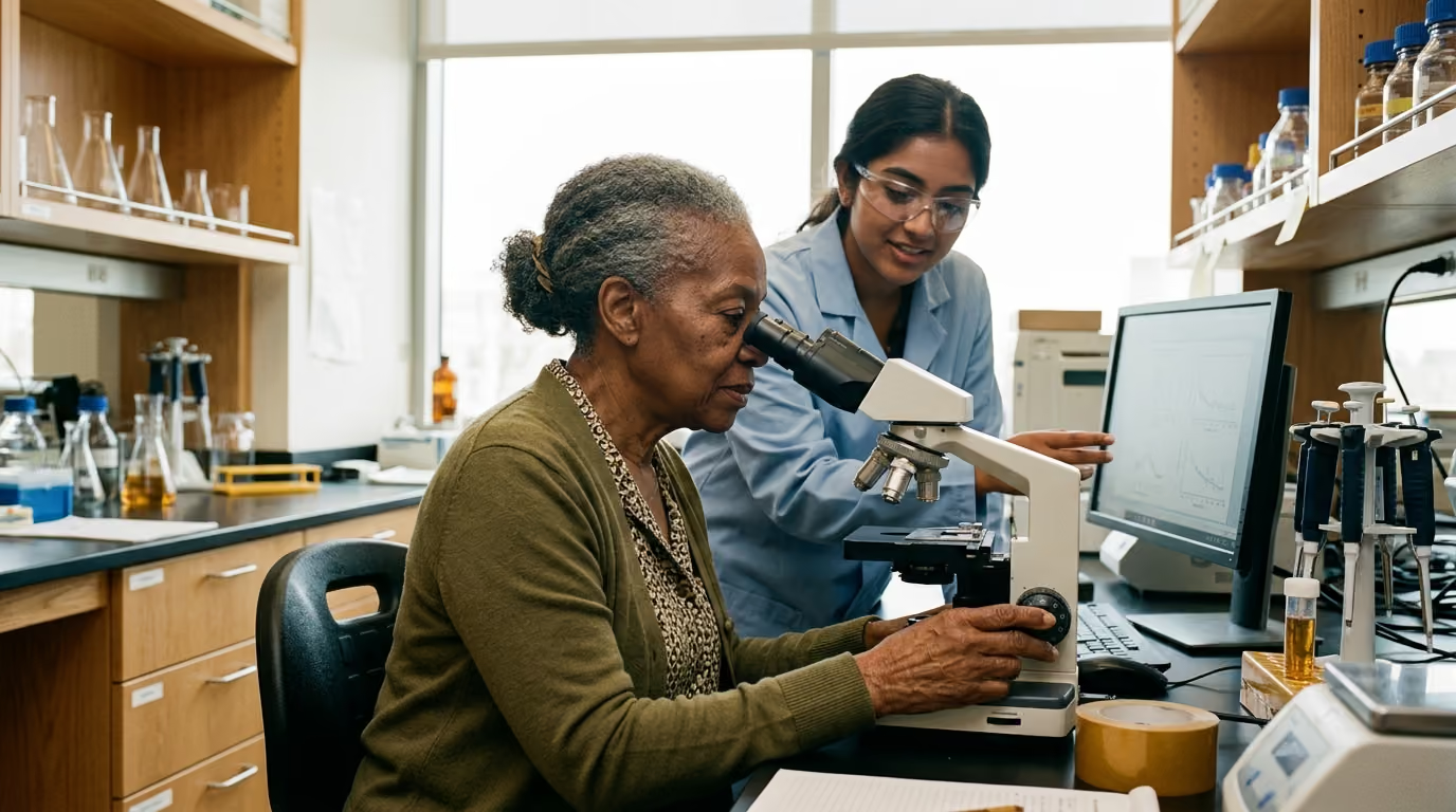

Metabolism slows down with age as peroxisomes lose their capacity to break down complex fatty acids and regulate cellular oxidative stress.





Many people search online to understand why cellular fat metabolism slows down with age. Most discussions focus almost entirely on mitochondria. Yet focusing solely on mitochondria leaves out an essential partner in cellular lipid metabolism. Peroxisomes are specialized organelles that carry out the initial breakdown of specific fatty acids, manage hydrogen peroxide levels, and communicate constantly with other cellular structures. This guide provides a definitive examination of peroxisomal biology, how these organelles alter their activity during aging, and what current scientific literature reveals about their role in metabolic health.
Recent scientific investigations demonstrate that cellular aging involves complex shifts in peroxisomal protein import, redox state, and metabolic capacity. In cultured human cells and aging rodent tissues, researchers observe specific declines in peroxisomal enzyme function alongside alterations in antioxidant defense. These findings come from distinct experimental systems, including single-cell yeast models, cultured human fibroblasts, and animal tissues. Because these experimental contexts differ substantially, findings from one model cannot be assumed to apply identically across all human tissues.
Understanding how peroxisomes function requires looking closely at three core factors. First is capacity, which reflects how much lipid breakdown and antioxidant processing an organelle can handle. Second is coordination, which describes how peroxisomes exchange metabolites and chemical signals with mitochondria and other organelles. Third is quality control, which involves how cells repair, replace, or degrade damaged peroxisomal components. Together, these factors shape how peroxisomes contribute to cellular health and metabolism over time.
Peroxisomes are single-membrane enclosed compartments found in almost all eukaryotic cells. They house more than fifty distinct enzymes dedicated to specialized metabolic pathways. While mitochondria handle the bulk of energy production from simpler fats, peroxisomes perform non-redundant biochemical tasks that mitochondria cannot execute alone.
The breakdown of fatty acids occurs through a repetitive chemical cycle termed beta-oxidation. In this process, fatty-acid chains are shortened step by step to produce smaller acyl-CoA molecules and acetyl-CoA. Mammalian cells divide this labor strategically between different compartments. Mitochondria process short-chain, medium-chain, and most long-chain fatty acids. Peroxisomes, by contrast, are required for the initial oxidation of very-long-chain fatty acids, branched-chain fatty acids, and specific lipid signaling molecules.
Very-long-chain fatty acids contain carbon chains of twenty-two or more carbon atoms. Because of their length and structure, these molecules cannot enter the mitochondrial matrix directly. Specialized transport proteins embedded in the peroxisomal membrane, known as ATP-binding cassette sub-family D transporters, import these large lipids into the peroxisome. Once inside, the fatty acids enter peroxisomal beta-oxidation to be shortened into medium-chain and short-chain segments.
The first step of peroxisomal beta-oxidation is catalyzed by the enzyme acyl-CoA oxidase. Acyl-CoA oxidase introduces a double bond into the fatty acyl-CoA substrate. During this reaction, the enzyme transfers electrons directly to molecular oxygen rather than to an electron transport chain. This enzymatic step produces an enoyl-CoA intermediate and generates hydrogen peroxide as an obligate byproduct.
After this initial oxidation, a multifunctional enzyme carries out subsequent hydration and dehydrogenation steps. Finally, a peroxisomal thiolase cleaves the molecule to yield acetyl-CoA and a shortened acyl-CoA chain. This cycle repeats until the fatty acid is reduced to an eight-carbon or six-carbon structure. These shortened products, along with acetyl-CoA, are then exported to the cytosol and mitochondria for full oxidation to carbon dioxide and water.
Through this coordinated division of labor, peroxisomes prevent the toxic accumulation of very-long-chain lipids while supplying shortened substrates for broader cellular and metabolic longevity pathways.
Peroxisomes process a distinct set of metabolic substrates that require specialized enzymatic handling:
The chemical reactions that occur inside peroxisomes make them unique among metabolic organelles. Because the rate-limiting step of peroxisomal beta-oxidation transfers electrons directly to oxygen, the organelle continuously generates hydrogen peroxide. Consequently, peroxisomes must maintain powerful antioxidant systems to manage this reactive oxygen species locally.
Peroxisomes are neither purely damaging nor purely protective. Instead, they act as dynamic redox nodes within the cell. The primary antioxidant enzyme inside peroxisomes is catalase. Catalase converts two molecules of hydrogen peroxide into water and molecular oxygen without requiring cellular energy. Peroxisomes also contain other protective enzymes, including copper-zinc superoxide dismutase, epoxide hydrolases, and peroxiredoxins.
In certain high-metabolism tissues, peroxisomal respiration accounts for a substantial fraction of total cellular oxygen consumption. In rodent liver tissue, researchers have reported that peroxisomal oxidases can account for up to thirty-five percent of total hydrogen peroxide production under standard physiological conditions. However, this measurement is specific to liver tissue and cannot be assumed to represent every cell type or organism.
Hydrogen peroxide functions as both a potential source of oxidative damage and an essential signaling molecule. At physiological levels, hydrogen peroxide diffuses through peroxisomal membrane channels to alter redox-sensitive cysteine residues on signaling proteins. This signaling helps regulate gene expression, stress adaptations, and lipid synthesis. When antioxidant defenses fail, excess hydrogen peroxide can leak into neighboring compartments and damage proteins, lipids, and nucleic acids.
Maintaining an appropriate balance between oxidant generation and antioxidant removal is essential. When peroxisomal catalase activity matches the rate of fatty acid oxidation, hydrogen peroxide remains confined to signaling roles. If peroxisomal protein import falters or catalase activity declines, this balance shifts, leading to local oxidative stress that can compromise cellular function.
Peroxisomes do not operate in isolation. They maintain close physical, metabolic, and signaling connections with other cellular structures, particularly mitochondria and the endoplasmic reticulum. This interplay is fundamental to the biology of aging and longevity science.
The cooperative relationship between peroxisomes and mitochondria occurs at two primary levels:
The metabolic output of peroxisomal beta-oxidation directly supports mitochondrial energy production. When peroxisomes shorten very-long-chain fatty acids, they produce acetyl-CoA, propionyl-CoA, and medium-chain acyl-CoAs. These shortened molecules leave the peroxisome through specialized carnitine-dependent and carnitine-independent export mechanisms.
Once imported into mitochondria, these metabolites feed directly into the tricarboxylic acid cycle. Acetyl-CoA combines with oxaloacetate to drive oxidative phosphorylation. Additionally, peroxisome-derived metabolites participate in anaplerotic reactions, which replenish tricarboxylic acid cycle intermediates that have been extracted for biosynthetic pathways. In organisms such as yeast, peroxisomal metabolism provides vital precursors that sustain mitochondrial respiration during nutrient shifts.
Beyond exchanging metabolic fuels, peroxisomes and mitochondria engage in continuous redox signaling. Experiments in cultured mammalian cells demonstrate that changing the antioxidant environment within peroxisomes produces measurable changes inside mitochondria.
In mouse embryonic fibroblasts engineered to lack functional catalase, researchers observed increased oxidation of mitochondrial redox reporters. When researchers restored catalase expression in these cells, the oxidation state of the mitochondrial reporters normalized. In related cellular experiments, acutely inhibiting catalase led to a statistically significant increase in mitochondrial oxidation.
Furthermore, when researchers induced oxidative stress exclusively within the peroxisomal matrix, the neighboring mitochondria exhibited altered redox states and excessive fragmentation. While these cell-culture models confirm a functional redox link, the exact mechanism remains under investigation. The effect may occur via direct diffusion of hydrogen peroxide, downstream lipid peroxidation products, or altered metabolic signaling molecules.
As cells age, their internal quality control systems and protein trafficking mechanisms experience measurable alterations. In peroxisomes, these changes are particularly evident in the efficiency of matrix protein import and the maintenance of internal redox balance.
Peroxisomes do not contain their own DNA. Every enzyme operating inside the peroxisome must be synthesized on cytosolic ribosomes and imported across the peroxisomal membrane. This transport relies on peroxisome targeting signals (PTS1 and PTS2) recognized by cytosolic receptor proteins called peroxins. The peroxin complex docks at the peroxisomal membrane, transfers the folded enzyme into the matrix, and recycles back to the cytosol.
Scientific investigations in cultured human diploid fibroblasts show that cellular senescence compromises this protein import machinery. As fibroblasts reach late passage states, the efficiency of importing PTS1-tagged and PTS2-tagged proteins declines. Catalase, which relies on a non-canonical PTS1 targeting sequence, appears especially vulnerable to these import defects. When catalase remains trapped in the cytosol, the peroxisomal matrix loses its primary hydrogen peroxide defense, leading to elevated intraperoxisomal oxidative stress.
A common misconception in cellular biology is assuming that a higher number of organelles always indicates superior metabolic function. Research using primary human fibroblasts demonstrates that organelle quantity does not equal organelle performance.
In late-passage senescent human fibroblasts, researchers observed a sharp increase in the total number of peroxisomes per cell. However, despite their greater numbers, most of these peroxisomes showed a reduced capacity to import targeted redox reporters. Using genetically encoded roGFP2 redox probes targeted to specific compartments, researchers quantified excitation ratios at 400 nm and 480 nm wavelengths to measure relative oxidation states.
In late-passage cells, the measured 400/480-nm excitation ratios were:
These assay-specific readouts revealed that while peroxisomes and mitochondria became significantly more oxidized in late-passage cells, the cytosolic compartment experienced the most pronounced shift toward oxidation.
The proliferation of dysfunctional peroxisomes in senescent cells likely reflects an impaired balance between organelle biogenesis and organelle removal. The selective degradation of damaged peroxisomes via autophagy, termed pexophagy, becomes less efficient during cellular senescence. As a result, damaged and import-incompetent peroxisomes accumulate within the cell instead of being recycled.
Data from whole-animal studies provide valuable context regarding how peroxisomal pathways behave in living tissues over time. Rodent models have been widely used to study enzymatic and structural shifts in liver, kidney, and brain tissues across the lifespan.
A consistent finding in rodent literature is the reduction of peroxisomal antioxidant defenses in aged liver tissue. Review literature indicates that catalase enzyme activity in liver samples from old rats and mice is reduced by approximately thirty to forty percent compared to young controls. This decline in enzymatic activity is accompanied by a reduced capacity to handle fatty acid oxidation substrates in aged hepatic tissue.
Researchers have also identified age-associated reductions in the activity of peroxisomal beta-oxidation enzymes, including acyl-CoA oxidase. In aging rat liver, studies describe reduced levels of the retinoid X receptor alpha (RXRα), which acts as a required binding partner for peroxisome proliferator-activated receptor alpha (PPARα). Because PPARα is the primary nuclear transcription factor driving peroxisomal enzyme synthesis, declining receptor levels offer a plausible mechanism for decreased beta-oxidation capacity.
However, these findings must be interpreted with appropriate scientific caution:
To evaluate longevity research accurately, one must separate observations in single-cell organisms from those in mammals. Model organisms provide vital mechanistic clues, but their biology does not always mirror human physiology.
The role of peroxisomal catalase presents a clear example of how experimental models can yield contrasting results. In the yeast Saccharomyces cerevisiae, peroxisomes are the sole site of fatty acid beta-oxidation. Yeast models demonstrate that peroxisomal lipid metabolism and mitochondrial communication influence stress responses and chronological lifespan.
Interestingly, several yeast studies show that genetic inactivation of catalase can extend chronological lifespan under specific growth conditions. Researchers hypothesize that the resulting low-level increase in hydrogen peroxide acts as an adaptive signal, activating downstream stress resistance pathways that enhance survival.
In mammalian cell cultures, however, catalase deficiency produces very different effects. When catalase is inhibited or genetically ablated in mouse or human fibroblasts, cells typically exhibit elevated mitochondrial oxidative stress, altered organelle morphology, and accelerated features of cellular senescence.
This dramatic difference highlights why researchers cannot assume that a biological mechanism in yeast will operate identically in human tissues. The cellular response to reactive oxygen species depends heavily on cell type, basal antioxidant capacity, growth conditions, and evolutionary adaptations.
Evaluating peroxisomal science requires distinguishing the type and scope of each experimental model:
In longevity research, distinguishing between validated clinical endpoints and surrogate biochemical markers is critical. A surrogate marker measures a biological molecule or cellular state, but it does not directly prove a change in total lifespan or clinical disease risk.
Understanding peroxisomal function involves several specialized laboratory and research measurements. Readers can explore broader diagnostic frameworks through resources on biological age testing and cellular health.
In clinical medicine, measuring plasma concentrations of very-long-chain fatty acids is the standard diagnostic test for peroxisomal disorders. Laboratories measure hexacosanoic acid (C26:0) and tetracosanoic acid (C24:0), along with the ratios of C24:0 to C22:0 and C26:0 to C22:0.
Elevated circulating levels of these saturated very-long-chain fatty acids indicate impaired peroxisomal beta-oxidation capacity. While these measurements are fully validated for diagnosing inherited metabolic diseases, their utility as general biomarkers of everyday human aging remains under ongoing scientific evaluation.
Plasmalogens are specialized ether phospholipids that make up a significant portion of cell membranes in the brain, heart, and immune cells. Because the initial steps of plasmalogen synthesis occur exclusively inside peroxisomes, circulating or tissue levels of plasmalogens reflect peroxisomal synthetic capacity.
Studies note that plasmalogen concentrations tend to decline in specific age-related conditions. However, blood plasmalogen levels can fluctuate based on diet, inflammation, and liver function, meaning they cannot serve as a standalone measure of biological age.
In laboratory research, genetically encoded fluorescent biosensors such as roGFP2 allow scientists to measure real-time oxidation states inside living cells. By targeting these probes to the peroxisomal matrix, cytosol, or mitochondria, researchers quantify dynamic changes in organelle redox status.
While these biosensors provide precise mechanistic data in cell culture and animal models, they cannot be deployed as routine clinical tests in living human patients.
While researchers continue to investigate specific pharmaceutical targets for peroxisomal function, several foundational lifestyle and nutritional factors support general cellular and organelle metabolism. Readers seeking a structured approach can review practical materials in our healthy aging resources collection.
optimal environment for cellular organelle maintenance.
A membrane-bound intracellular organelle containing enzymes for fatty acid beta-oxidation, ether lipid synthesis, and hydrogen peroxide metabolism.
A fatty acid possessing a carbon chain of twenty-two or more carbon atoms, requiring initial metabolic breakdown within peroxisomes before mitochondrial entry.
The rate-limiting enzyme in peroxisomal beta-oxidation that catalyzes the initial oxidation step, transferring electrons to molecular oxygen to yield hydrogen peroxide.
A heme-containing antioxidant enzyme abundant within peroxisomes that rapidly breaks down hydrogen peroxide into molecular oxygen and water.
A family of proteins essential for peroxisome biogenesis, membrane assembly, and the import of matrix enzymes from the cytosol.
A specialized form of selective autophagy dedicated to the recognition, engulfment, and lysosomal degradation of damaged or surplus peroxisomes.
The biochemical process of replenishing metabolic intermediate molecules in the tricarboxylic acid cycle that were consumed during biosynthetic pathways.
A class of ether-linked phospholipids abundant in brain and cardiac cell membranes whose initial synthetic reactions occur exclusively inside peroxisomes.
A nuclear receptor protein that forms a heterodimer with peroxisome proliferator-activated receptor alpha (PPARα) to regulate the transcription of peroxisomal and mitochondrial fatty acid oxidation genes.
The balance between oxidized and reduced chemical species within a specific cellular compartment, governing oxidative stress levels and redox-sensitive signaling pathways.
Stay current with research on aging biology, biomarkers, nutrition, therapeutics, peptides and longevity technology. AgeAmaze reports what the evidence shows, where uncertainty remains and which claims still need stronger data.


Follow AgeAmaze for careful reporting on what longevity science can show today and what still needs stronger evidence.
read the Blog