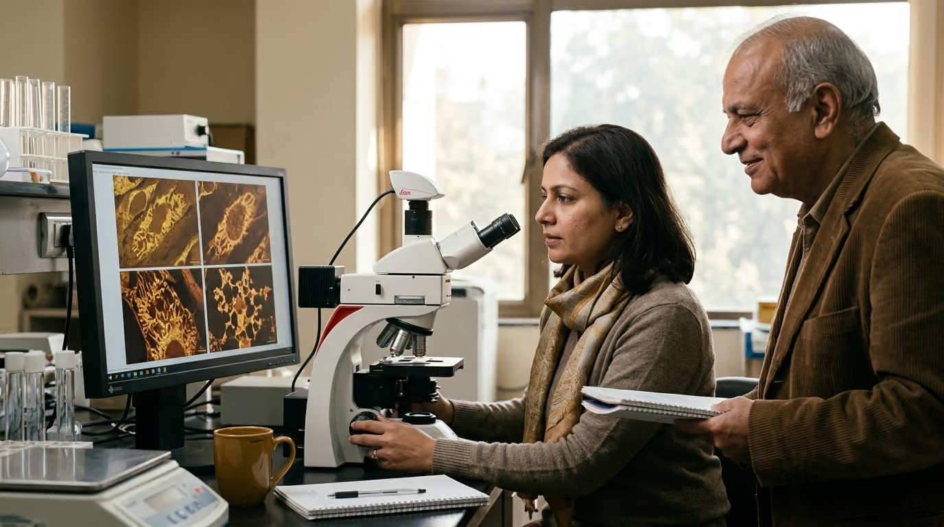

A detailed perspective on cellular decline highlights how mitochondrial dysfunction disrupts cellular signaling, triggers chronic inflammation, and accelerates the biological aging process.





Mitochondrial aging is not a simple story of cellular batteries running out of power. It represents a complex shift in how cells balance energy production, chemical signaling, structural maintenance, and stress responses over time. Understanding this biology requires moving past outdated metaphors to examine how these dynamic organelles coordinate cellular survival and how those regulatory networks change across a lifespan.
Researchers who study cellular health and metabolism research view mitochondria as central communication hubs. These structures couple nutrient oxidation to energy storage while managing calcium movement, redox signaling, and programmed cell death. When mitochondrial systems falter, the consequences ripple across tissues. Yet scientific evidence shows that these changes vary widely across different cell types and organs.
This comprehensive guide analyzes the current scientific understanding of mitochondrial aging. It evaluates the biochemical mechanisms of energy conversion, the maintenance systems that preserve organelle quality, and the competing scientific models that attempt to explain why mitochondrial performance alters with age. It also examines human clinical evidence, highlights the critical difference between biomarker shifts and true lifespan extension, and outlines the methodological challenges that shape modern geroscience.
The standard description of mitochondria as cellular powerhouses captures only a fraction of their physiological responsibility. Mitochondria generate adenosine triphosphate through oxidative phosphorylation, but they also serve as vital sensory and signaling platforms. They continuously monitor nutrient availability, oxygen tension, and cellular stress, sending signals that alter nuclear gene expression and dictate cell fate.
Energy conversion occurs across the inner mitochondrial membrane through the electron transport chain. Complexes I through IV transfer electrons derived from carbohydrates and fats to molecular oxygen. This transfer pumps protons from the matrix into the intermembrane space, establishing an electrochemical proton gradient. Complex V, known as ATP synthase, harnesses this potential energy to phosphorylate adenosine diphosphate into ATP.
Beyond generating ATP, the electron transport chain produces reactive oxygen species as natural metabolic byproducts. Electron leakage, primarily at Complexes I and III, partially reduces oxygen to generate superoxide. Cells rapidly convert superoxide
into hydrogen peroxide, which acts as a diffusible signaling molecule. In healthy cells, transient pulses of hydrogen peroxide regulate protein phosphorylation, activate protective transcription factors, and adjust metabolic output.
Mitochondria also control intracellular calcium balance. By sequestering and releasing calcium ions, they shape cytosolic calcium signals that regulate muscle contraction, neurotransmission, and enzyme activation. Sustained calcium overload inside the mitochondrial matrix, however, can trigger the opening of the mitochondrial permeability transition pore. This pore opening collapses the membrane potential, arrests ATP production, and releases cytochrome c into the cytoplasm to initiate apoptosis.
Mitochondria do not exist as isolated, static capsules. They form dynamic, interconnected networks that continuously divide, fuse, and remodel. This continuous structural adaptation allows the organelle network to share components, isolate damaged sections, and maintain metabolic efficiency. Mitochondrial homeostasis relies on a coordinated balance among four primary quality-control mechanisms.
Biogenesis is the generation of new mitochondrial mass and protein components. This process requires coordinated expression from two distinct genomes: the nuclear genome and mitochondrial DNA. The transcriptional coactivator peroxisome proliferator-activated receptor gamma coactivator 1-alpha serves as the master regulator. It activates nuclear respiratory factors, which subsequently stimulate mitochondrial transcription factor A to drive the replication and transcription of mitochondrial DNA.
Dynamics describe the physical remodeling of mitochondrial networks through fission and fusion. Mitofusins 1 and 2, along with optic atrophy 1, mediate the fusion of outer and inner mitochondrial membranes. Fusion allows partially damaged mitochondria to complement each other by mixing matrix contents, structural proteins, and undamaged genomes. Conversely, dynamin-related protein 1 drives fission, dividing networks to separate depolarized or heavily damaged fragments for targeted degradation.
Mitophagy is the specialized autophagy pathway that isolates and degrades dysfunctional mitochondria inside lysosomes. When an individual mitochondrion loses its inner membrane potential, the kinase PINK1 stabilizes on its outer membrane. Accumulated PINK1 recruits and phosphorylates the E3 ubiquitin ligase Parkin, which coats damaged outer membrane proteins with ubiquitin chains. Autophagy receptors recognize these markers and recruit the organelle into an autophagosome, preventing damaged structures from generating excessive reactive species.
The mitochondrial unfolded protein response represents a retrograde signaling pathway activated when unfolded or misfolded proteins accumulate inside the organelle. When matrix proteases cannot keep pace with misfolded proteins, specific peptide fragments travel to the cytoplasm and nucleus. This signal triggers the transcription of nuclear genes that encode mitochondrial chaperones and quality-control proteases. These newly synthesized chaperones enter the organelle to restore protein homeostasis and preserve respiratory function.
Scientific investigations reveal clear age-associated patterns across diverse tissues, but these changes are neither uniform nor identical across the body. In many tissues, aging correlates with lower mitochondrial oxidative capacity, altered organelle architecture, and an accumulation of oxidative macromolecular modifications. However, researchers emphasize that structural content and functional respiratory performance represent distinct biological variables.
A tissue can maintain a normal quantity of mitochondria while suffering from diminished respiration per organelle. Alternatively, a tissue might experience a reduction in total mitochondrial volume while the remaining organelles maintain intact respiratory efficiency. Conflating these two properties leads to incorrect conclusions about tissue decline. Measuring both properties simultaneously is necessary to evaluate the true metabolic status of aging cells.
These alterations manifest in distinct ways across organs:
Age-associated mitochondrial alterations do not advance as a single synchronized wave throughout the body. An individual may demonstrate preserved mitochondrial respiratory capacity in cardiac tissue while exhibiting significant declines in sedentary skeletal muscle. Recognizing tissue specificity prevents oversimplified generalizations about biological aging.
Geroscience does not present a single, universally accepted model for mitochondrial aging. Instead, researchers evaluate several competing, overlapping mechanisms. Each framework explains specific experimental observations, yet none accounts for every feature of tissue aging across all species.
The classical Mitochondrial Free Radical Theory of Aging proposed a vicious cycle. The hypothesis stated that electron transport produces reactive oxygen species that damage mitochondrial DNA and proteins. This damage causes respiratory chain dysfunction, which generates even higher amounts of reactive species, accelerating cellular destruction.
Modern investigations have largely overturned this circular model. Experimental studies demonstrate that genetically increasing antioxidant enzymes in animal models does not consistently extend lifespan. Furthermore, moderate elevations in reactive oxygen species often correlate with longer lifespan in model organisms.
These findings led to the concept of mitohormesis. This model establishes that low-level, transient mitochondrial stress generates reactive signals that activate endogenous cytoprotective defenses. While chronic, overwhelming oxidative stress damages cellular components, physiological amounts of reactive species are necessary for healthy adaptation.
Mitochondrial DNA resides near the electron transport chain and lacks histone protection, making it susceptible to replication errors and oxidative modifications. Each cell contains hundreds to thousands of copies of mitochondrial DNA, a state known as polyplasmy. When mutations arise, they create heteroplasmy, where mutated genomes coexist with wild-type sequences inside the same cell.
A mutated genome does not immediately impair cellular respiration. Experimental models indicate that cells generally tolerate mutated genomes until they cross a critical threshold, often estimated between 60% and 90% of total copies. Once heteroplasmy exceeds this threshold, the cell develops measurable respiratory chain deficiencies.
Through non-uniform replication during cell division, mutated copies can expand clonally over time. This clonal expansion creates a mosaic pattern in aging tissues, where severely deficient cells sit directly adjacent to entirely normal cells.
Another prominent model attributes organelle decline to the progressive failure of quality-control networks. As cells age, the efficiency of both biogenesis and mitophagy decreases. Reduced biogenesis prevents the timely replacement of worn components, while sluggish mitophagy allows depolarized, damaged organelles to persist inside the cytoplasm.
This persistence disrupts cellular metabolism. Damaged mitochondria consume ATP in reverse to maintain their membrane potential, release pro-inflammatory molecules, and generate aberrant calcium signals. When quality control fails, the cell becomes trapped in a state where neither repair nor disposal functions properly.
A fourth perspective focuses on the contraction of critical metabolic cofactor pools, particularly nicotinamide adenine dinucleotide. The oxidized form, NAD+, serves as an essential electron acceptor in glycolysis and the tricarboxylic acid cycle. It also acts as a required substrate for sirtuins, a family of deacetylases that regulate mitochondrial biogenesis, antioxidant defenses, and DNA repair.
With advancing age, increased activity of NAD-consuming enzymes, such as CD38 and poly(ADP-ribose) polymerases, can lower intracellular NAD+ availability. This reduction impairs the activity of SIRT1 and SIRT3, blunting PGC-1alpha activation and weakening mitochondrial stress responses. Although this pathway provides valuable insights into metabolic flexibility, manipulating cofactor levels in healthy humans has not been shown to extend lifespan.
Mitochondrial dysfunction does not remain confined within the individual organelle. Severe or persistent mitochondrial distress triggers signaling cascades that alter cellular identity and contribute to systemic, low-grade inflammation. This phenomenon forms an essential bridge within biology of aging and longevity science resources.
Cellular senescence is a state of durable cell-cycle arrest accompanied by characteristic metabolic and secretory changes. While severe DNA damage or telomere shortening triggers classical senescence, metabolic impairment inside mitochondria initiates a distinct variant known as Mitochondrial Dysfunction-Associated Senescence.
MiDAS develops when mitochondrial respiratory failure or severe NAD+ depletion prevents the cell from maintaining necessary energetic fluxes. Cells undergoing MiDAS arrest their growth through p53 and p21 pathways without exhibiting typical markers of nuclear DNA damage. Furthermore, the secretory profile of MiDAS cells differs significantly from classical senescence.
Cells in MiDAS secrete specific pro-inflammatory cytokines while lacking many of the matrix-remodeling enzymes seen in genotoxic senescence. This distinction proves that senescence is a heterogeneous collection of cellular states rather than a single uniform program.
Mitochondria evolved from ancient alpha-proteobacterial endosymbionts, meaning their internal components resemble bacterial structures. Under normal conditions, these components remain sequestered inside the inner and outer membranes. When severe stress compromises membrane integrity, mitochondria release these molecules into the cytoplasm or extracellular fluid, where the immune system recognizes them as danger signals.
These innate immune cascades create a state of persistent, sterile inflammation termed inflammaging. This inflammation damages adjacent healthy cells, interferes with stem cell maintenance, and degrades extracellular matrix structures across aging tissues.
The degree to which age-related mitochondrial changes are fixed versus modifiable remains a critical area of investigation within cellular and metabolic longevity science. Disentangling chronological age from physical inactivity is notoriously difficult in human studies, particularly when evaluating skeletal muscle.
Cross-sectional studies in humans illustrate this challenge. Sedentary older adults frequently demonstrate lower mitochondrial volume, reduced oxidative enzyme activities, and impaired respiration compared to younger controls. However, older adults who engage in lifelong aerobic training display mitochondrial profiles that closely resemble those of younger active individuals.
A comprehensive review of human skeletal muscle demonstrates that active older subjects maintain a 48.9% greater mitochondrial volume density than their sedentary age-matched peers. Furthermore, a structured four-month aerobic exercise intervention in older adults elevated mitochondrial protein synthesis, boosted oxidative enzyme activities, and stimulated biogenesis pathways to levels comparable to younger adults.
A systematic review of randomized controlled trials confirms that structured training produces robust improvements in:
These adaptations prove that human skeletal muscle retains remarkable plasticity into advanced age. However, these positive adaptations must be interpreted with scientific rigor. Demonstrating that exercise improves mitochondrial respiration in muscle does not prove that it halts intrinsic biological aging across all organs or extends maximum human lifespan. Exercise enhances functional capacity and metabolic healthspan, but it does not make the organelle immune to time-dependent molecular changes.
Evaluating mitochondrial longevity research requires an objective understanding of the experimental limitations and translation gaps that characterize the field. Many widely publicized concepts stem from isolated cell models or short-lived model organisms that do not fully mirror human aging biology.
Much of the foundational mechanistic literature derives from model organisms like Caenorhabditis elegans, Drosophila melanogaster, or laboratory mice. These species possess distinct metabolic rates, mitochondrial genetics, and lifespan determinants compared to humans. An intervention that extends lifespan in a roundworm by inducing mitohormesis may produce no meaningful benefit, or even cause toxicity, in a complex human physiology.
Traditional laboratory techniques often measure biomarkers in whole-tissue homogenates. This approach averages biochemical signals across millions of heterogeneous cells. Because mitochondrial DNA mutations and respiratory defects spread in a mosaic distribution, bulk assays can mask severe, localized dysfunction in a subset of critical cells. Emerging single-cell sequencing and spatial respirometry methods are beginning to address this gap, but substantial methodological hurdles remain.
A critical limitation in modern longevity literature is the premature conflation of biomarker improvements with biological rejuvenation. Modulating a surrogate readout, such as increasing mitochondrial DNA copy number or elevating tissue NAD+ levels, does not establish that biological aging has been reversed or that clinical longevity has increased.
Scientific reviews explicitly emphasize that no pharmacological intervention targeting mitochondria has been proven to slow biological aging or extend maximum lifespan in healthy human populations.
Assessing mitochondrial health involves a diverse set of laboratory biomarkers. Each metric captures a specific aspect of organelle structure, abundance, or biochemical function, yet none provides a complete picture on its own. Understanding the exact scope and limitations of these markers is essential when interpreting biological age testing methods.
Citrate synthase is a key pace-making enzyme located in the mitochondrial matrix that catalyzes the first step of the Krebs cycle. Its activity level correlates closely with total mitochondrial volume and structural mass within a tissue sample. However, high citrate synthase activity indicates only that mitochondrial material is present; it does not confirm that the respiratory chain is coupling oxygen consumption to ATP synthesis efficiently.
Respirometry measures the rate of oxygen consumption in permeabilized cells or isolated organelles across various substrate-uncoupler-inhibitor titration protocols. It assesses:
Mitochondrial DNA copy number quantifies the average number of mitochondrial genomes relative to nuclear DNA in a biological sample. While frequently evaluated in blood cells, its utility as an aging biomarker remains complex. A higher copy number can reflect robust biogenesis, but it can also represent a compensatory response where failing cells overproduce defective genomes to offset functional deficiencies.
This ratio evaluates the redox state and metabolic health of the cytoplasm and mitochondrial matrix. A high NAD+/NADH balance supports efficient glycolysis, fatty acid oxidation, and sirtuin-mediated deacetylase activity. Reductions in this ratio signal metabolic congestion, impaired electron transport throughput, or excessive cofactor consumption by repair enzymes.
When cells experience necrotic death, severe trauma, or unchecked senescence, they release fragmented mitochondrial DNA into the bloodstream. Elevated plasma levels of cell-free mitochondrial DNA serve as a validated biomarker of systemic inflammation, tissue damage, and cellular stress. It functions as an indicator of cellular disruption rather than a direct readout of organelle bioenergetics.
No. Reactive oxygen species are essential signaling molecules required for normal cellular function, immune responses, and metabolic regulation. While chronic, excessive oxidative stress damages lipids, proteins, and nucleic acids, transient low-level bursts of reactive species stimulate protective adaptations through mitohormesis. Suppressing all reactive species with high-dose antioxidants can disrupt these beneficial signaling pathways.
Scientific evidence does not support the claim that broad antioxidant supplementation reverses mitochondrial aging or extends human lifespan. In many clinical trials, non-targeted antioxidant supplements failed to improve metabolic health or reduce age-related pathology, and in some cases interfered with exercise-induced adaptations. Mitochondrial quality depends on dynamic homeostatic regulation, quality control, and functional stress signaling rather than simply neutralizing free radicals.
Current tests measuring mitochondrial DNA copy number or mutations provide only partial snapshots of cellular state. These metrics vary widely across different tissues, fluctuate with physical activity levels, and can rise compensatorily during metabolic stress. While informative within research frameworks, an isolated mitochondrial DNA readout cannot establish an accurate, comprehensive biological age for an individual.
Structured physical exercise is the most robust, clinically validated intervention for enhancing mitochondrial health in human skeletal muscle. Aerobic and resistance training stimulate biogenesis, optimize network dynamics, promote mitophagy, and increase oxidative enzyme capacity. While various pharmaceutical agents targeting metabolic pathways remain under active investigation, none has demonstrated the broad systemic benefits and safety profile of regular physical activity in humans.
Stay current with research on aging biology, biomarkers, nutrition, therapeutics, peptides and longevity technology. AgeAmaze reports what the evidence shows, where uncertainty remains and which claims still need stronger data.


Follow AgeAmaze for careful reporting on what longevity science can show today and what still needs stronger evidence.
read the Blog




